Clinical Presentation
65 year old woman with a history of a previous right total hip replacement performed 14 years ago. Now complaining of chronic pain in the right groin and inner thigh with a clunking sensation during walking. The orthopaedic surgeon was concerned about loosening of the hip replacement, so a bone SPECT-CT scan of the hips was performed.
Planar Imaging Findings
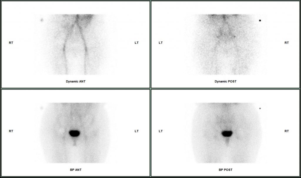
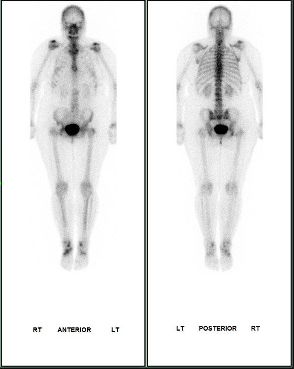
Dynamic and blood pool phase planar imaging was unremarkable. Whole body delayed imaging shows focal increased uptake around the right ischial tuberosity.


Key SPECT-CT findings
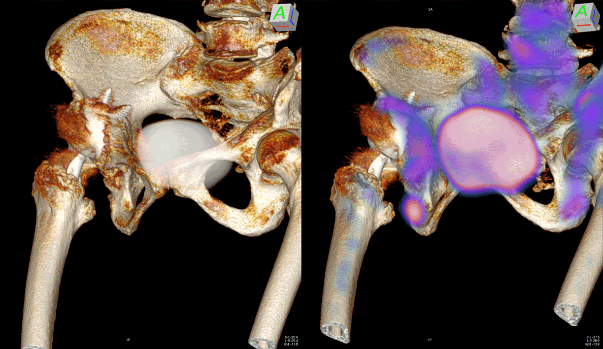
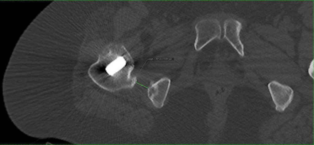
SPECT-CT through the pelvis and hips reveals focal increased uptake in the right ischial tuberosity. Whilst the initial impression may be of periostitis related to hamstring origin tendinopathy, the location of the uptake is on the lateral surface of the ischial tuberosity. In addition, there is cortical irregularity with cystic changes in the bone at the site of uptake. On reviewing the ischiofemoral space, the ischiofemoral distance was noted to be reduced compared to the left side.
There are no features of loosening of either the femoral or acetabular components of the hip replacement.
There is increased uptake in the left sacroiliac joint due to altered biomechanical stress.


Diagnosis
Ischiofemoral impingement of the right hip
Ischiofemoral impingement
Ischiofemoral impingement is a rare cause of pain in the hip due to soft tissue impingement between the ischial tuberosity and the lesser trochanter. The mechanical constriction of the soft tissues, particularly the quadratus femoris, causes the pain.
Clinical features are pain in the buttock, groin and inside of the thigh, radiating to the knee. Patients can be symptomatic more several months or years. A snapping or clunking sensation can be experienced by patients during walking as the lesser trochanter bypasses the ischial tuberosity. Patients sometimes describe worsening of symptoms during pronounced extension of the hip, e.g running or taking large steps.
There several causes for narrowing of the ischiofemoral space:
- Congenital changes such as coxa valga and developmental dysplasia of the hip
- Osteoarthritis with superomedial migration of the femur
- Proximal femoral fractures
- Osteochondroma of the femur or pelvis
- Following total hip replacement
Imaging Findings
MRI reveals soft tissue oedema of the quadratus femoris muscle. In chronic cases there may be fatty atrophy of the quadratus femoris muscle and tears of the hamstring origin tendons.
On SPECT-CT the ischiofemoral distance is easiest to measure. In a study by Torriani et al. a cut off of <17mm for ischiofemoral distance gave a sensitivity of 83% and specificity of 82%.
The differential diagnosis includes tear of the quadratus femoris, adductor muscle tear or tendinitis of hamstring origin.
SPECT-CT allows evaluation of the bone turnover that occurs secondary to the chronic repetitive stress from soft tissue impingement. In our case, the combination of the increased uptake on bone scintigraphy and reduced ischiofemoral space confirms the diagnosis.


